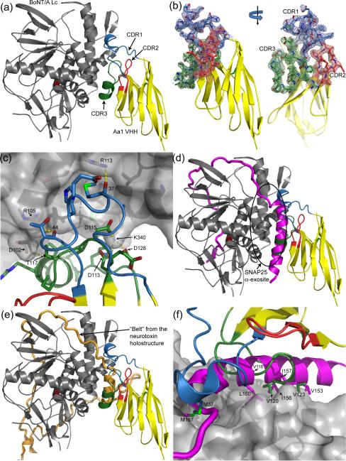
Figure 5. Structure of the BoNT/A Lc - Aa1 VHH complex.
(A) BoNT/A Lc endopeptidase in gray complexed with the Aa1 VHH fragment in yellow with the CDR1, CDR2, and CDR3 regions colored blue, red, and green, respectively. The catalytic zinc is depicted as a red sphere in all figures. (B) Surface representation of the BoNT/A Lc highlighting the Aa1 VHH binding site. Six hydrogen bonds between the Lc and the Aa1 VHH fragment are indicated with yellow dashes. (C) The SNAP25 natural substrate colored in magenta from PDB code 1XTG superimposed onto the BoNT/A Lc – Aa1 VHH complex. The α-helical portion of SNAP25 that binds to the BoNT/A Lc α-exosite coincides with the α-helical tips of CDR1 and CDR3. (D) The same superposition from panel (C) highlighting the amino acid conservation between the SNAP25 α-exosite binding region and the Aa1 VHH fragment. (E) The “belt” from the BoNT holostructure colored orange (from PDB code 3BTA), superimposed onto the BoNT/A Lc – Aa1 VHH complex. The α-helical tips of CDR1 and CDR3 coincide with an α-helical portion of the “belt” in a fashion similar to the SNAP25 / VHH superposition shown in panel (C).

