
Figure 1.
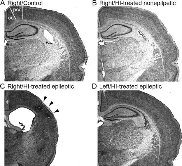
Unilateral infarct after ligation of the right carotid artery at postnatal day 7. A–C, Coronal sections from control animal (A), HI-treated animal without an ischemic lesion (B), and an HI-treated rat with a clear ischemic lesion (C, D). The rat brains were stained with cresyl violet (i.e., Nissl stain). Right hemispheres in A and B are comparable, in contrast to C, which shows the parasagittal infarct (arrowheads) and related cortical atrophy and an enlarged lateral ventricle. The contralateral (left) hemisphere (D) is shown from the same section as in C and illustrates that the left hemisphere was not lesioned. cc, Cingulate cortex; pcc, paracingulate cortex.