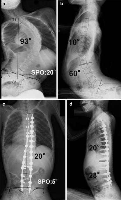
Fig. 3.
a, b A 13-year-old boy with DMD. Anteroposterior radiograph shows severe scoliosis of 93° with significant pelvic obliquity of 20°. Thoracic hypokyphosis and lumbar hyperlordosis were present. c, d All-screw constructs and fusion to L5 were performed. Postoperative sitting views show significant coronal curve correction of 20° with normalization of sagittal plane. Pelvic obliquity improved to 5°

