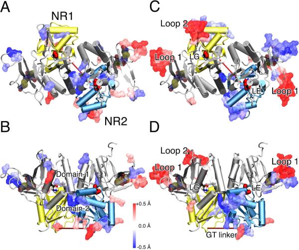
Figure 5.
Difference of backbone flexibility between the xenon and control systems for the ligand-binding domain of the NMDA receptor in the closed-cleft conformation (cartoon) is highlighted using a color gradient: from the most increased flexibility (red) to the most decreased flexibility (blue). (A) Top and (B) site views of the RMSF changes in the presence and absence of xenon over the last 5-ns simulations. (C) Top and (D) site views of the changes based on GNM analysis in the mean square fluctuation of the three lowest motions in the systems with and without xenon atoms. The glycine (LG) and glutamate (LE) agonists are presented in solid surface. Domain-1 is colored silver in both subunits. Domain-2 is colored in yellow in NR1 and light blue in NR2. The disulphide bonds in Loop 1 are highlighted in transparent surface and colored by atom type. Xenon atoms were not included in the GNM network, as explained in the Supporting Information by Fig. S6.

