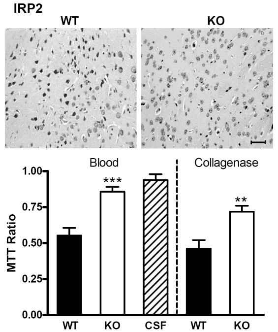
Fig 6.
IRP2 knockout increases striatal cell viability after ICH. Representative photomicrographs of sections from wild-type (WT) and IRP2 knockout (KO) striata 72 h after striatal blood injection, stained with hematoxylin and eosin. The left border of each photo is 350 μm from the injection site. Scale bar = 50 μm. Bar graph represents mean striatal cell viability three days after striatal blood or collagenase injection (±S.E.M, 5-11/condition), as assessed by cellular conversion of MTT to formazan. All values are normalized to those in contralateral striata (= 1.0) to yield MTT signal ratio. WT and KO mice injected with artificial CSF had similar values, so results were combined. ***P< 0.001, **P< 0.01 compared with the ratio in WT mice injected with blood or collagenase.

