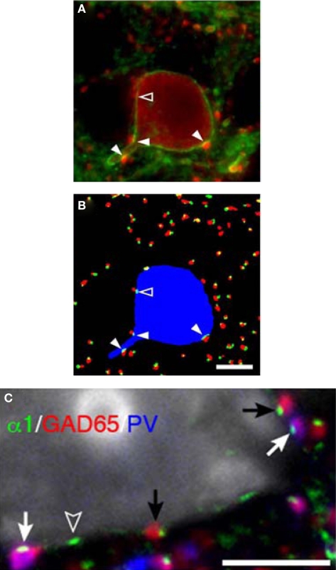
Figure 6.
Parvalbumin-positive basket cell boutons apposing GABAA α1 subunits in the plasma membrane of neurons. (A) In a single confocal z-plane GABAA α1 (green) is seen in the plasma membrane of a PV (red) neuron in a tissue section. (B) Color segmentation masks of the image in (A) which represents the intracellular PV (blue), PV boutons (red) and GABAA α1 clusters (green). The different masks were made using different parameters for our iterative approach. Only the red and green object masks that partially overlap, and thus correspond to PV puncta and α1 clusters that are directly apposed, are shown. The arrowheads point to α1 clusters that are within the PV neuron and are directly apposed by PV puncta. Note that in (A) several PV puncta appear to juxtapose the soma near the open arrowhead. However, in this z-plane only one is directly apposed to an α1 cluster (open arrowhead). (C) A single confocal z-plane of a tissue section was labeled for GABAA α1 subunit (green), GAD65 (red), PV (blue), and Nissl substance (gray). Note GAD65 and PV dual-labeled (purple) boutons (white arrows) and GAD65 labeled boutons (black arrows) in apposition to GABAA α1 subunits in the plasma membrane of a pyramidal cell. The open arrowhead indicates an α1 cluster for which the apposed bouton is below the plane of focus. All Bars = 5 μm.

