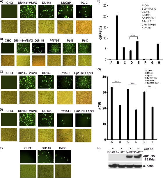
FIG. 4.
XMRV pseudovirus infectivity in prostate cancer cell lines (A), immortalized prostate fibroblast cell lines (B), epithelial cell lines with or without Xpr1 transfection (C), smooth muscle cell lines (D) with or without Xpr1 transfection, and primary epithelial cells (PrEC) (E). Representative images in the fluorescence and bright fields were captured at ×20 magnification. The experiments were carried out twice, and each time the experiments were performed in triplicate. (F) The average levels of GFP expression in infected cells were quantified by FACS analysis. Xpr1 expression levels were determined by quantitative real-time RT-PCR (G) and Western blotting (H). Asterisks indicate statistical significance (P < 0.001).

