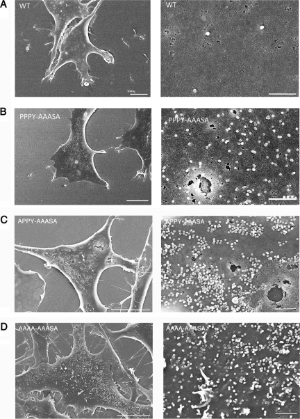
FIG. 9.
Budding morphology of late domain mutants. Shown are representative scanning electron micrographs of DF1 cells transiently transfected with the wild type (A) and the PPPY-AAASA (B), APPY-AAASA (C), and AAAA-AAASA (D) mutants. The cells shown had similar levels of GFP fluorescence. The scale bars are 10 μm in the left images and 1 μm in the right images.

