FIGURE 1.
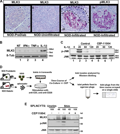
MLK3 is activated in leukocyte-infiltrated islets of NOD mice, by IL-1β in Min6 cells, and in SICC. A, pancreatic sections from prediabetic (panel i) and diabetic NOD mice (panels ii–iv) were stained for MLK3 (panels i–iii) and phospho-MLK3 (panel iv) as described. Leukocyte infiltration is outlined. Bar, 20 μm. B, Western blotting of extracts (60 μg of protein) from Min6 cells treated for 20 min with 10 nm tumor necrosis factor-α, 40 ng/ml interferon-γ, or 20 ng/ml IL-1β using anti-MLK3 antibodies and β-tubulin used as a control. C, Western blots for time-dependent JNK activation from Min6 cells treated with IL-1β (20 ng/ml) in the absence (lanes 2–4) or presence of MLK3 inhibitor CEP11004 (lanes 5–7). D, schematic of SICC. E, Western blots for total MLK3 and phospho-JNK (pJNK) from of SICC islets co-cultured with increasing numbers of splenocytes (4- and 10-fold for 30 min) in the absence (lanes 1–3) or presence of CEP11004 (lanes 4 and 5). Total JNK levels serve as control. NT, not treated.

