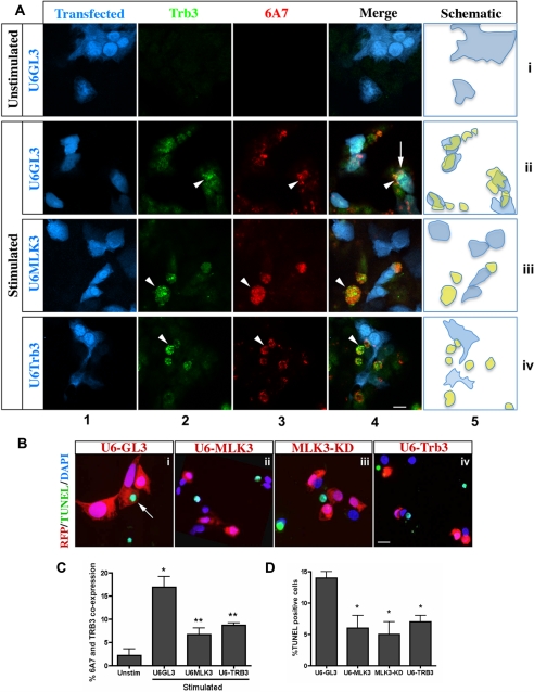
FIGURE 4.
Cytokine-induced BAX conformational change, and apoptosis requires MLK3 and TRB3. A, Min6 cells were cotransfected with shRNA constructs U6-GL3 (row ii), U6-MLK3 (row iii), or U6-TRB3 (row iv), with RFP (pseudocolored blue, column 1) to track transfected cells. Induction of endogenous TRB3 (green, column 2) and its colocalization with BAX-6A7 (column 3, pseudocolored red) was detected following an 8-h treatment with unstimulated (row i) or stimulated (rows ii–iv) medium. Column 4 shows a merge of RFP (pseudocolored blue) marker to track shRNA uptake, BAX-6A7, and TRB3 signals. Arrowheads, colocalization of TRB3 and BAX-6A7 in the absence of shRNA; arrows, TRB3 and BAX-6A7 in the presence of shRNA control U6-GL3. Bar, 10 μm. A schematic representation of the overlapping expression of shRNA (blue) and TRB3/BAX-6A7 double positive cells (yellow) is shown in column 5. B, TUNEL analysis of Min6 cells co-transfected with RFP reporter and either U6-GL3 (panel i), U6-MLK3 (panel ii), MLK3-KD (panel iii), or U6-TRB3 (panel iv) were treated with stimulated medium for 24 h. Arrows, TUNEL, shRNA/RFP double positive. Bar, 1 μm. C, quantification (means ± S.D. (error bars) of three independent experiments) of shRNA-positive cells co-expressing BAX-6A7 and TRB3 (*, p < 0.001 versus unstimulated media; **, p < 0.001 versus U6-GL3, as tested by ANOVA followed by Bonferroni post hoc test). D, TUNEL quantification of RFP positive cells (mean ± S.D. of three experiments; *, p < 0.001 versus U6-GL3, as tested by ANOVA followed by Bonferroni post hoc test).

