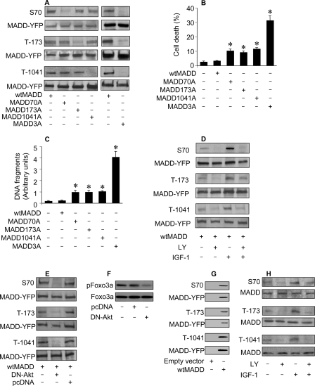
FIGURE 2.
In vivo phosphorylation of MADD. A, detection of MADD phosphorylation using specific anti-phospho antibodies is shown. HEK293 cells were transfected with various cDNA constructs, and 36 h later the cells were harvested for immunoblotting using the anti-phospho-Ser-70, anti-phospho-Thr-173, or anti-phospho-Thr-1041 antibodies or an anti-YFP antibody to detect exogenous MADD. B, shown is an analysis of cell fate. HEK293 cells were transfected with various MADD constructs. Cell death was determined 48 h after transfection by trypan blue exclusion. *, p < 0.05 versus control. C, measurement of apoptosis using Cell death detection enzyme-linked immunosorbent assay is shown. Cells were treated as described for B. *, p < 0.05 versus control. D, induction, or inhibition of MADD phosphorylation is shown. HEK293 cells were transfected with wtMADD, and 24 h later they were cultured in serum-free medium for 20 h. Subsequently, they were treated with LY294002 (LY, 10 μm) for 1 h and/or IGF-1 (150 ng/ml) for 20 min. The cell lysates were immunoblotted and probed with anti-phospho-specific antibodies or an anti-YFP antibody. E, DN-Akt inhibits MADD phosphorylation. HEK293 cells were co-transfected with wtMADD along with dominant negative Akt (DN-Akt) or an empty vector pcDNA3.1 (pcDNA). Immunoblots were probed with indicated antibodies. F, DN-Akt could reduce Foxo3a phosphorylation levels. HeLa cells were transfected with DN-Akt or an empty vector pcDNA3.1 (pcDNA). Immunoblots were probed with the anti-Foxo3a antibody or the anti-phospho Foxo3a (T32) antibody. G, exogenous MADD is expressed and phosphorylated. HEK293 cells were transfected with either a empty vector is a vector containing YFP-fused MADD. Expression and phosphorylation of exogenous MADD were analyzed by immunoblotting. H, phosphorylation of endogenous MADD is shown. HeLa cells were serum-starved for 20 h and treated with LY (10 μm) for 1 h and/or IGF-1 (150 ng/ml) for 20 min. Immunoblot was probed with the indicated antibodies.

