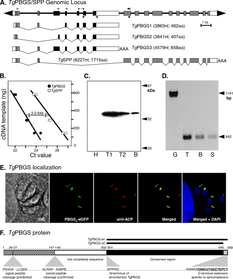
FIGURE 1.
TgPBGS organization, expression, and localization. A, 23 kb spanning the T. gondii PBGS/SPP locus on chromosome III. Exons 1 and 2 encode a bipartite apicoplast-targeting signal (signal sequence + plastid-targeting domain; hatched boxes), fused to mature coding sequences for TgPBGS (exons 3–7; black boxes) or TgSPP (exons 8–17; gray boxes). The white boxes indicate 5′- and 3′-untranslated regions (precise polyadenylation sites have not been mapped for TgPBGS1 and -2). The open arrowheads indicate primers used to define the 5′-end of TgPBGS and TgSPP transcripts by reverse transcription-PCR or RACE, bars indicate fragments amplified for quantitative PCR analysis of mRNA abundance, and closed arrowheads indicate primers used for assessing TgPBGS expression in different parasite stages. Three distinct transcripts including PBGS sequences were identified. B, quantitation of TgPBGS and TgSPP transcripts by quantitative real-time PCR using gene-specific primer pairs (supplemental Table S1); TgPBGS was amplified above the Ct threshold ∼2 cycles earlier than TgSPP, corresponding to a 3.5-fold difference in mRNA abundance (replicate samples at three different dilutions). C, Western blotting of parasite cell lysates detects the 56-kDa PBGS3 protein. H, primary human foreskin fibrobast host cell lysate; T1, 5 × 10⁶ T. gondii tachyzoites; T2, 106 tachyzoites; B, 400 T. gondii bradyzoite cysts (purified from a chronically infected mouse brain). D, a 442-bp PCR product specific for TgPBGS3 mRNA was detected in tachyzoites (T), bradyzoites (B), and sporozoites (S). G, amplification of genomic DNA using the same primers yields a 1.1-kb product. E, subcellular localization of TgPBGS in transgenic parasites expressing the N-terminal leader (394 amino acids) fused to eGFP (green). The apicoplast (red) was detected by immunostaining for T. gondii acyl carrier protein (ACP). 4′,6-diamidino-2-phenylindole (DAPI) (blue) stains the apicoplast and both parasite and host cell nuclei. DIC, differential interference contrast. F, schematic diagram of TgPBGS protein features showing the bipartite plastid-targeting region (hatched), predicted signal peptide, and experimentally determined transit peptide cleavage sites and the portions expressed as recombinant proteins. aa, amino acids; nt, nucleotides.

