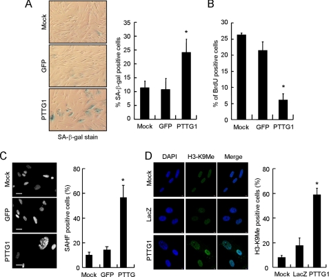
FIGURE 2.
Senescent phenotype observed in PTTG1-expressing normal human fibroblasts. IMR90 cells were transduced with adenoviruses carrying PTTG1 or GFP and then cultured at 37 °C for 4 days. A, the transduced cells were analyzed for SA-β-gal activity staining. Photographs of the X-gal-stained cells are shown. Quantification of the SA-β-gal positive-stained cells was conducted (right). Results were obtained from the average of three independent experiments. An asterisk indicates p < 0.05 (p = 0.0172). B, the virus-transduced cells were cultured in medium containing BrdUrd for 4 h. After labeling, the cells were fixed and stained with anti-BrdUrd antibody. The percentage of BrdUrd-positive cells is presented. Results were obtained from the average of three independent experiments. An asterisk indicates p < 0.05 (p = 0.000081). C, the virus-transduced cells were stained with DAPI and then visualized under a fluorescence microscope (left). The heterochromatin foci were quantified from three independent experiments (right). An asterisk indicates p < 0.05 (p = 0.003). D, shown are confocal images of indirect immunofluorescence of histone H3 methylation on lysine 9 (H3-K9Me) in mock control or LacZ- or PTTG1-expressed IMR90 cells. The DNA was also stained by DAPI. Quantification of the H3-K9Me foci-positive cells was conducted (right). An asterisk indicates p < 0.05 (p = 0.0003).

