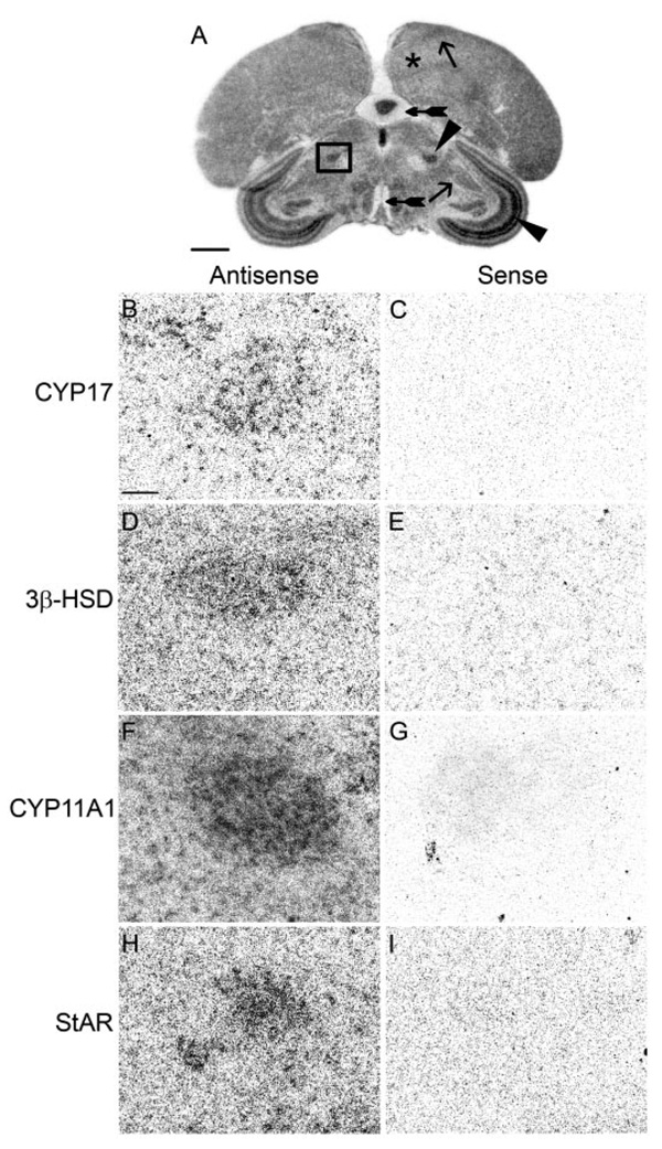
FIG. 4.
The medial nucleus of the spiriform complex, a sensory integration area, expresses CYP17, 3β-HSD, CYP11A1, and StAR. A, Low-magnification image of a film image showing position of the SpM (darkly stained nucleus in box) in an adult male section hybridized for StAR. B–I, High-magnification dark-field images of area in box. B, D, F, and H, Emulsion-dipped sections hybridized with antisense configured probes for CYP17, 3β-HSD, CYP11A1, and StAR, respectively. Adjacent sections hybridized with sense probes (C, E, G, and I) do not show label. Areas representative of low (+), medium (++), and high (+++) hybridization levels are indicated. The telencephalon with low hybridization is marked with an asterisk; the ICo/MLd and HVC determined to have medium hybridization are marked with an arrow; the SpM and SGC, areas of high levels of hybridization, are marked with an arrowhead; and the nerve tract of the III cranial nerve and the molecular layer of the cerebellum, which did not show hybridization, are indicated with tailed arrows. Scale bar (A), 1 mm; (B), 100 µm (B–I).

