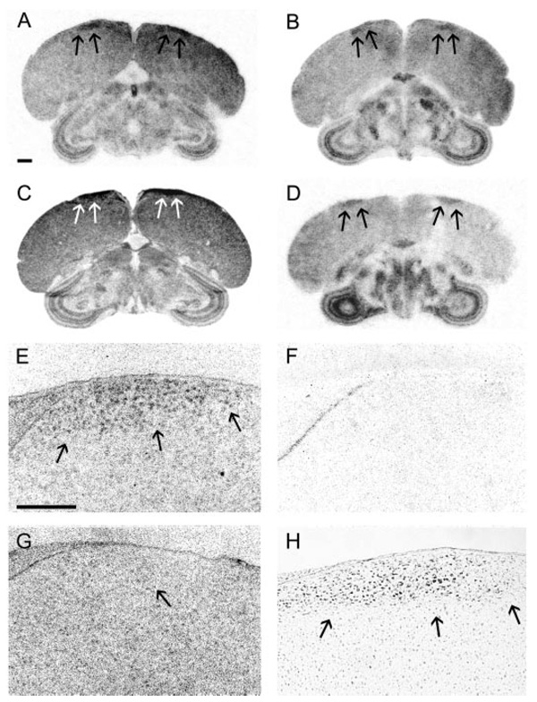
FIG. 6.
StAR, CYP11A1, 3β-HSD, and CYP17 are expressed in male HVC. A–D, Film images show position of HVC hybridized for StAR (A), CYP11A1 (B), 3β-HSD (C), and CYP17 (D) in adult male brain. Arrows indicate position of HVC. E–G, Inverted dark-field images of emulsion-dipped slides hybridized for CYP11A1 show specific cellular hybridization in male HVC hybridized with antisense (E) but not sense-configured probes (F). HVC label is not evident in adult female hybridized with antisense (G) configured probes. Arrow (G) indicates analogous region to HVC in the female. High-magnification image of a nissl-stained section shows the cellular anatomy of male HVC (H). Scale bars (A), 1 mm (A–D) and (E) 300 µm (E-H).

