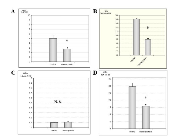
Figure 1.

Quantification of IL-6, TNF-α, IL-1β and TLR5 mRNA in jejune samples. Figure 1A and 1B illustrate in a bar diagrams that IL-6 and TNF-α mRNA expression increased markedly in control group compared with mannoprotein. Figure 1C shows IL-1β mRNA expression in both groups represented as bars diagram (n = 9 control group, n = 8 mannoprotein group). No statistical differences were observed between both groups. Finally, figure 1D shows the results for TLR5 gene expression in both groups with augmented levels in the control group compared with mannoprotein. Error bars represent the standard deviations. * Significant at p > 0.05 compared with control.
