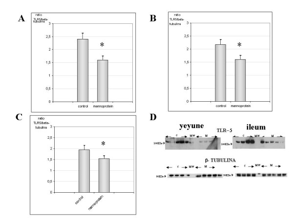
Figure 4.

Quantification of TLR5 protein levels in yeyune, Ileum and colon samples., Figure 4A, 4B and 4C show the levels of TLR5 protein expression in both groups for yeyune, ileum and colon respectively. In all figures an augmented expression of TLR5 is observed in the control group compared with mannoprotein (n = 9 control group, n = 8 mannoprotein group). These differences were statistically significant. Figure 4D shows a representative Western-blot picture of TLR5 and β-tubuline in yeyune (left) and Ileum (right) for both groups controls (designed as C) and mannoprotein (designed as M). Error bars represent the standard deviations. * Significant at p < 0.05 compared with control.
