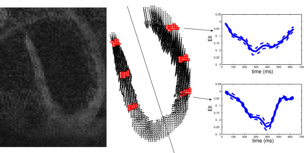
Figure 4.
A sample image of DENSE (left) and corresponding data acquisition points (middle) are illustrated on the left ventricle wall in the CMR long-axis slice of a healthy human heart. The middle panel also shows the 2D projection of the motion for these material points at the time of maximum contraction (360 ms after QRS). The six regions determined with red dots are at 30%, 55% and 80% of the distance from the apex to the aortic valve in which the comparisons of Tables 4 and 5 have been performed. (Right) Mean value of ELL as a function of time, shown in two regions of the free wall; the solid plot lines show ELL averaged over the points, while the dashed lines show mean ± std over the same group of points.

