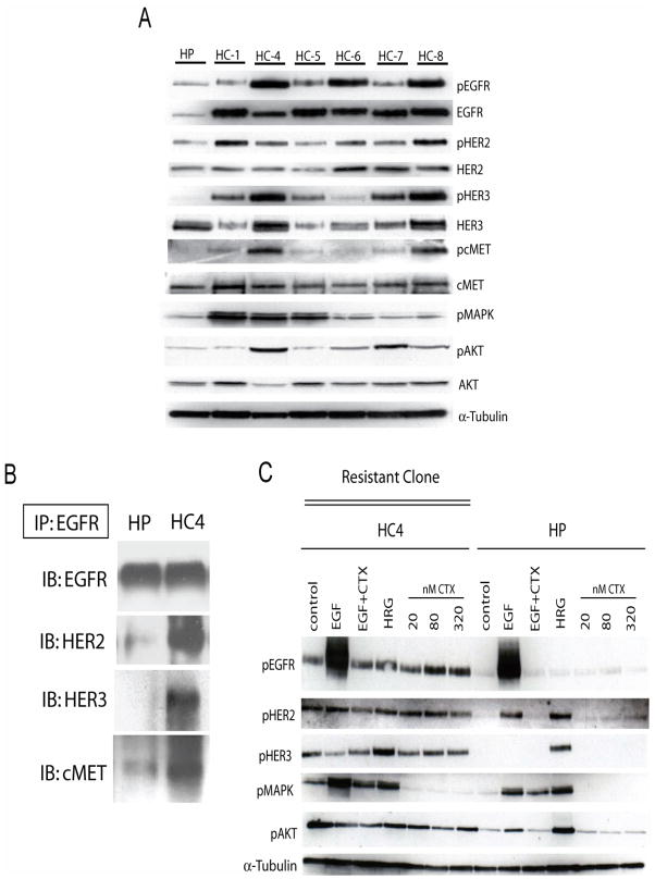
Figure 2. EGFR is upregulated in cetuximab-resistant cells.
A: Characterization of expression of HER family members and down stream Akt and MAPK in cetuximab-resistant clones (HC1, HC4-HC8). Protein was collected and fractionated by SDS-PAGE followed by immunoblotting for the indicated proteins. α-tubulin was used as loading control. HP; cetuximab-sensitive parental line, HC; cetuximab-resistant clones.
B: EGFR has increased association with HER2, HER3, and cMET in cetuximab-resistant cells. Cetuximab-resistant cells were harvested and EGFR was immunoprecipitated from the cetuximab-resistant clone HC4 and the parental HP cells with an anti-EGFR antibody. The immunoprecipitates were fractionated on SDS-PAGE followed by immunoblotting for the indicated proteins. HP; cetuximab-sensitive parental line, HC4; cetuximab-resistant clone.
C: Cetuximab treatment does not modulate EGFR phosphorylation in cetuximab-resistant clone (HC4). Parental cells (HP) and cetuximab-resistant clone (HC4) were treated with vehicle (control) or increasing concentrations of cetuximab (CTX) for 24 hours. Stimulation was with EGF (10ng/ml) and HRG (5μM) for 45 minutes. Protein was collected and fractionated by SDS-PAGE followed by immunoblotting for the indicated proteins. α-tubulin was used as a loading control.

