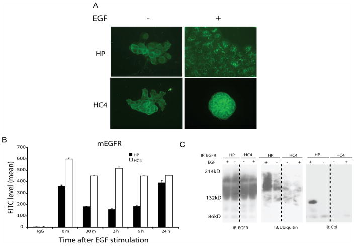
Figure 3. Cetuximab-resistant cells have increased EGFR plasma membrane expression.
A: EGFR is internalized following EGF stimulation in parental (HP), but not in cetuximab-resistant cells (HC4). Internalization of EGFR was determined by immunofluorescent staining as described in “Experimental Procedures” using FITC-conjugated anti-EGFR antibody 45 min following EGF (25 ng/ml) stimulation. Internalized EGFR was indicated by the accumulation of intracellular endocytic vesicles staining for EGFR.
B: Cell surface EGFR is decreased following EGF stimulation in parental (HP), but not in cetuximab-resistant cells (HC4) The levels of EGFR on the cell surface were monitored following 0~24 hr of EGF (25 ng/ml) stimulation by flow cytometry as described in “Experimental Procedures”. The numbers represent mean fluorescence intensity of anti-EGFR staining after subtracting IgG background fluorescence.
C: Lack of c-Cbl recruitment and EGFR ubiquitination in cetuximab-resistant cells following EGF stimulation. Cetuximab-resistant (HC4) and parental (HP) cells were either untreated (-) or treated with EGF (25 ng/ml) for 45 min. Thereafter, cell lysates were prepared and analyzed by immunoprecipitation (IP) and immunoblotting (IB) with the indicated antibodies.

