
Figure 5. Rolling and immobilized leukocytes after infusion of NO releasing nanoparticles (NO-np) and control nanoparticles (Control-np).
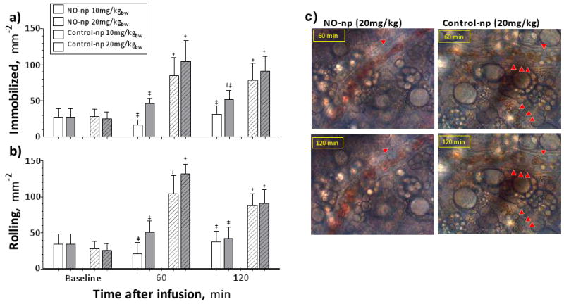
a) Immobilized leukocytes after infusion of 10 (white bars) and 20 (gray bars) mg/kg of NO-np (no pattern) and Control-np (pattern upward diagonal). 6 animals were used in each group, 6-8 venules were selected in each animal. †, P<0.05 to Baseline; ‡, P<0.05 to Control-np at same concentration. b) Rolling leukocytes after infusion of NO-np and Control-np. Rolling leukocytes were quantified in the same animals and locations as Immobilized. †, P<0.05 to Baseline; ‡, P<0.05 to Control-np at same concentration. c) Immobilized and rolling leukocytes after infusion of 20 mg/kg of NO-np and Control-np. Red arrows point to immobilized leukocytes on the images, other leukocytes in the images are rolling of flowing.