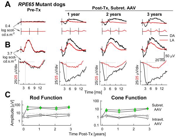
Figure 12.
Long term restoration of vision in RPE65-mutant dogs by gene therapy. (A) ERGs evoked by standard white flashes in the right eye of an RPE65-mutant dog before treatment (Pre-Tx) and over a 3-year interval after treatment. Flashes presented under dark-adapted (DA) and light-adapted (LA) conditions. DA traces are single waveforms, LA traces are averages obtained at repetition frequencies of 1 (top) and 29 Hz (bottom). Black vertical lines show the timing of the flashes. (B) ERG photoresponses evoked with white flashes of high energy over the same 3-year interval in the same eye as in A. Waveforms displayed as in A. (C) Two eyes with subretinal AAV-RPE65 (green symbols) show stable level of partial restoration of retinal rod and cone function, whereas two eyes with intravitreal AAV-RPE65 (gray symbols) show amplitudes similar to those of untreated eyes. Horizontal dashed lines represent the upper limit (mean + 3 SD) of the respective measurement in the group of control RPE65-mutant affected eyes, which had not received treatment. Modified from Acland et al., 2005.

