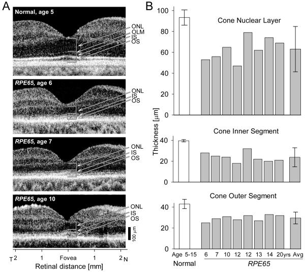
Figure 2.
Foveal cone morphology of young RPE65-LCA patients. (A) Cross-sectional scans along the horizontal meridian of the central retina of a normal child (upper panel) and three children with LCA due to RPE65 mutations (lower panels). Arrows and brackets indicate ONL, outer nuclear layer; OLM, outer limiting membrane; IS, inner segment; OS, outer segment. (B) Mean foveal ONL, IS and OS thickness in a group of young subjects with normal vision (ages 5–15 years) and in eight young patients with RPE65–LCA (ages 6–20 years). Mean values for the parameters from the RPE65–LCA group are also shown (Avg). Error bars represent +/−2 SD. Reprinted from Maeda et al., 2009, by permission from Oxford University Press.

