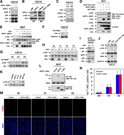
Figure 5.
Cdo and APPL1 are required for Akt activation in myoblast differentiation. (A) Confluent cultures of C2C12 cells stably expressing APPL1 RNAi were immunoblotted recognizing antibodies to pp38, p38, Cdo, JLP, or APPL1 to show depletion of APPL1. (B) C2C12 cells stably expressing APPL1 or control pBabePuro (pBP) expression vectors, and APPL1 RNAi or control (pSuper) vectors were cultured to confluency and induced to differentiate for 1 d (D1) or for 2 d (D2), respectively. Cell lysates were immunoblotted with antibodies to p-Akt, Akt, or APPL1 to show the level of overexpression or knockdown of APPL1 protein. (C) C2C12 cells were cultured either to 50% confluency (LD) or 100% confluency (HD) in growth medium, and lysates were immunoblotted with antibodies to Cdo, APPL1, p-Akt, or Akt. (D) 293T cells were transiently transfected with GST-Cdointra, HA-Akt, SRT-APPL1, or control pcDNA (−) expression vector as indicated. Lysates were pulled down with glutathione-Sepharose beads and analyzed by immunoblotting with antibodies to Cdo, SRT, HA, and p-Akt. (E) 293T cells were transiently cotransfected with 2.5 μg of Cdo, and varying amounts of APPL1 (0.25, 0.5, 1.25, and 2.5 μg) constructs, and 2.5 μg of Cdo, APPL1, or control, pcDNA (−) vector-transfected cells serve as controls. Lysates were analyzed after 48 h by immunoblotting with antibodies to p-Akt, Akt, SRT, or Cdo. (F) C2C12 cells stably transfected with control pSuper or Cdo RNAi were immunoblotted with antibodies to Cdo or Cadherin as a loading control. (G) Cell lines shown in F were cultured to near confluency and induced to differentiate for up to 2 d. Lysates were analyzed with antibodies to p-Akt or Akt. (H) Primary myoblasts isolated from Cdo+/+ or Cdo−/− mice were harvested at various differentiation time points. Lysates were immunoblotted with antibodies against p-Akt, Akt, or Cdo as control for the genotype of Cdo. (I) Extracts of hindlimb muscles from the control wild-type or Cdo−/− embryos at E15.5 were analyzed by immunoblotting with antibodies to p-Akt, Akt, pp38, p38, or Cdo as control. (J) C2C12 cells stably expressing Cdo, Cdo mutant (Δ1035-1160), or control pBP were cultured in DM for 2 d, and lysates were immunoblotted with antibodies to Cdo, p-Akt, Akt, MHC (indicative of differentiation response), or Cadherin as a loading control. p-Akt, Akt, MHC, and Cadherin loading control signals were quantified by densitometry; ratio reported under each lane in arbitrary units with C2C12/Cdo was set to 1. (K) Lysates of C2C12 cells stably expressing APPL1, APPL1 mutants, or control pcDNA (cell lines from Figure 4C) were immunoblotted with antibodies against p-Akt or Akt. (L) 293T cells were transiently transfected with HA-tagged APPL1, and lysates were subjected to coimmunoprecipitation with an antibody to Akt followed by Western blotting with antibodies to Akt and HA. (M) Control and APPL1 RNAi C2C12 cells from various differentiation time points were analyzed for cell death by TUNEL staining (red), followed by DAPI (blue) staining. (N) Quantification of TUNEL/DAPI staining shown in M. Values represent means of triplicate determinations ±1 SD. The experiment was repeated twice with similar results.

