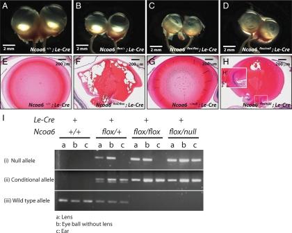
Figure 10.
Deletion of Ncoa6 in prospective lens ectoderm led to microphthalmia and cataract. (A–D) Eyeball morphology of four genotypes: Ncoa6+/+; Le-Cre, Ncoa6flox/+; Le-Cre, Ncoa6flox/flox; Le-Cre, and Ncoa6flox/null; Le-Cre at ∼3 mo old. (E–H) H&E staining of 3-mo-old lenses from Ncoa6+/+; Le-Cre and Ncoa6flox/flox; Le-Cre mice, and 1-mo-old lenses from Ncoa6+/null; Le-Cre and Ncoa6flox/null; Le-Cre mice. Scale bar is shown in each panel. (I) To analyze the deletion efficiency of the floxed allele, lens, eyeball (without lens), and ear tissues from Ncoa6+/+; Le-Cre, Ncoa6flox/+; Le-Cre, Ncoa6flox/flox; Le-Cre, and Ncoa6flox/null; Le-Cre mice were used to extract genomic DNA for genotyping. Analysis of ear tissue confirmed the tissue-specific expression of Le-Cre. In Ncoa6+/+; Le-Cre tissues, only the WT allele was detected. Most Ncoa6flox/+; Le-Cre tissues showed the WT, conditional and null alleles except ear tissues in which no null allele is detected. In Ncoa6flox/flox; Le-Cre mice, the null allele is detected in the lens; however, the deletion is incomplete as in the Ncoa6flox/null; Le-Cre lens.

