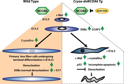
Figure 12.
Summary diagram of lens fiber cell differentiation and the role of NCOA6 in the denucleation process. In WT lens (left side of the diagram), up-regulation of c-Maf and crystallins is essential for lens fiber cell differentiation. Their terminal differentiation requires orchestrated degradation of all subcellular organelles to avoid light scattering. Nuclear degradation is a relatively lengthy process (3–4 d in mouse; Bassnett, 2009). In Cryaa-dnNCOA6 transgenic lenses (right side of the diagram), p53-dependent and p53-independent proapoptotic processes become evident as early as at E13.5 due to the generation of CCAP3 followed by the appearance of DSBs monitored via γ-H2AX–specific antibodies. Although abundant TUNEL signals are found at E16.5, the nuclear degradation is not properly executed and abnormally differentiated lens fibers, with significantly reduced expression of c-Maf and γ-crystallins and retained nuclei, are preserved in the transgenic lenses.

