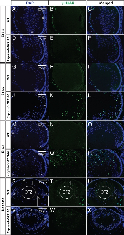
Figure 6.
Identification of DNA double-strand breaks (DSBs) through γ-H2AX immunofluorescence in WT and Cryaa-dnNCOA6 lens fiber cells. γ-H2AX antibody was used to label DSBs generated through DNA damage or normal denucleation process. DSBs are detected as early as E13.5 in the transgenic lens fiber cell compartment (D–F) and are absent in the WT lens (A–C). The number of DSBs increased in E14.5 and E16.5 transgenic lens fiber cells (J–L and P–R), whereas WT controls are negative (G–I and M–O). In the neonatal WT lens, nuclei of lens fiber cells at the margin of the OFZ display strong γ-H2AX staining (S–U and S′–U′). The neonatal transgenic lens exhibits dispersed DSBs staining in fiber cells and the absence of OFZ (V–X). The OFZ is indicated by dashed circle. Nuclei (blue signal) were stained with DAPI. Mesenchymal cells and hyaloid vascular structure surrounding the lens often generate nonspecific signals, which do not interfere with our observations in the lens fiber cell compartment. Lens epithelium, LE; transitional zone, TZ. Scale bar is shown in A, D, G, J, M, P, S, and V.

