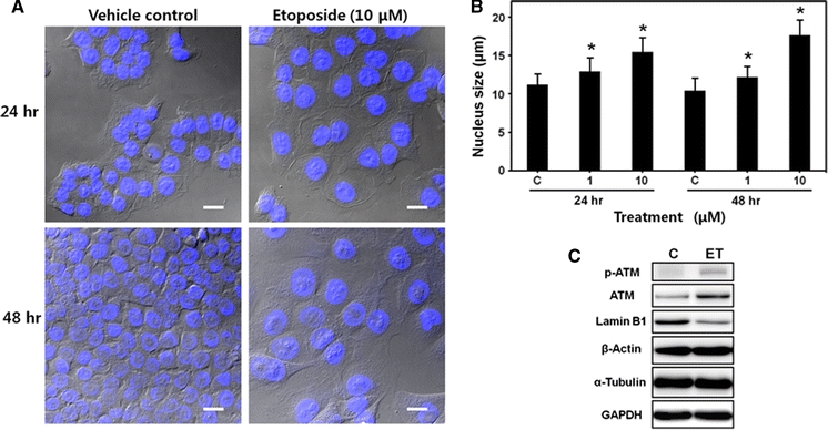
Fig. 3.

Enlargement of cell and nucleus by etoposide treatment in HCT116 cells. a Confocal microscope images of HCT116 cells treated with etoposide (10 μM) for 24 or 48 h. Cells were fixed and stained with DAPI. Differential interference contrast (DIC) images were superimposed on DAPI fluorescence images (bar = 20 μm). b Measurement of nuclear size. HCT116 cells treated with etoposide (0–10 μM) for 24 or 48 h, fixed, and stained with PI. Nuclei size was determined by fluorescence microscopy and the circle measurement algorithm of the microscope software. Each bar represents the mean ± SD (n = 100). * P < 0.001 compared to vehicle control, C. c Expressions of ATM, lamin B1, β-actin, α-tubulin, and GAPDH in HCT116 cells determined by Western blot analysis. ATM phosphorylation was also measured. Cells were treated with etoposide (ET, 10 μM) for 24 h. C vehicle control. Representative data are shown from two independent experiments
