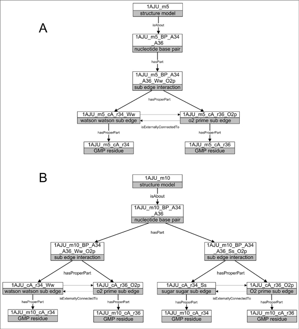
Figure 2.
RKB nucleotide base pairs with varying sub-edge interactions. Illustration of the RDF-based representation of molecular structure obtained from PDB files and from structure feature analysis of MC-Annotate. (A) Structure model 5 of PDB 1AJU is about a nucleotide base pair that is composed of a sub-edge interaction between the Watson-Watson sub-edge of the guanine residue at position 34 of chain A and the O2’ sub-edge of the guanine residue at position 36 of chain A. (B) Structure model 10 of PDB 1AJU is about a nucleotide base pair between guanine residue at position 34 in chain A and guanine residue at position 36 in chain A, which is composed of two sub-edge interactions – a Watson-Watson sub-edge and O2’ sub-edge, as well as a Sugar-sugar sub-edge and the O2’ sub-edge.

