
Fig. 5.
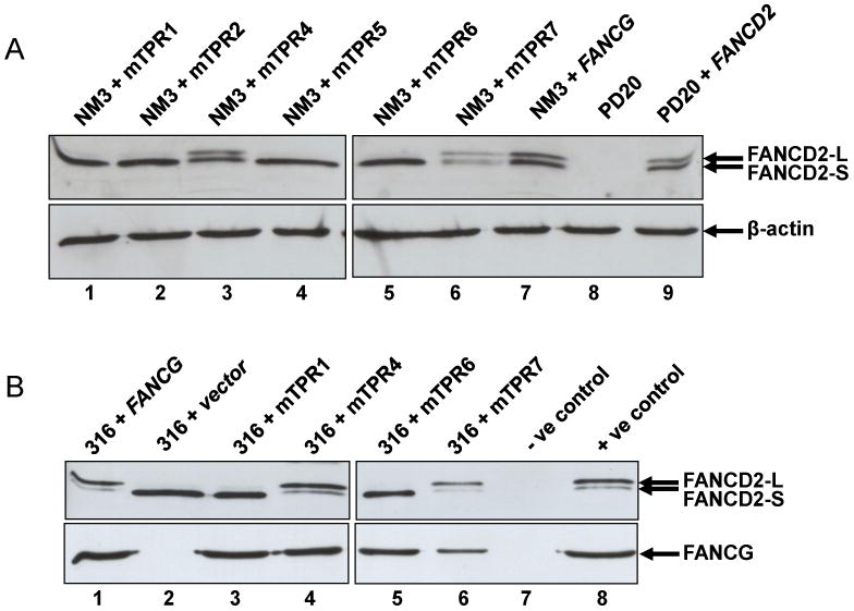
Expression of FANCD2 in NM3 and EUFA316 cell lines with mutated TPR motifs. Whole cell extracts were prepared from NM3 or EUFA316 cells treated with 50 nM MMC for 18 h. (A) NM3+mTPR1, +TPR2, +TPR5, and +TPR6 lack significant expression of the monoubiquitylated isoform (FANCD2-L), whilst NM3+TPR4 and +TPR7 and NM3+FANCG express both monoubiquitylated and non-ubiquitylated (FANCD2-S) isoforms. Human PD20 (FA-D2) cells and a cDNA-corrected counterpart (PD20+FANCD2) were used as negative and positive controls for FANCD2 detection by the antibody (lanes 8 & 9 respectively). β-actin was used as a loading control. (B) EUFA316+vector, +mTPR1, and +TPR6 lack significant expression of FANCD2-L, whilst EUFA316+mTPR4, +TPR7 and EUFA316+FANCG express both isoforms. Expression of FANCG in the same cell extracts are shown in the lower panel. As negative and positive controls for the detection of FANCD2 by the antibody, PD20 and PD20+FANCG were used (lanes 7 & 8 upper panel), whilst NM3 and NM3+FANCG were used as controls for FANCG (lanes 7 & 8 lower panel).