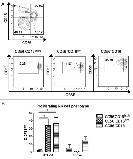
Figure 3.
Phenotype of proliferating NK cells after seven days' culture ex vivo. (A) NK cells were first identified as viable CD3− lymphocytes based on FSC/SSC, aqua amine-reactive dye, and anti-CD3 staining. The NK cells were then gated into three subsets based on CD56 and CD16 expression (upper) and CFSE dilution was measured within each subset (lower). (B) For each of the three defined NK cell phenotypes, proliferating cells were measured in HTLV-1 infected and normal control subjects. A minimum of 30,000 live lymphocytes was collected for each subject, and cell subsets with at least 50 gated events were included for analysis of proliferation (%CFSElow). Error bars represent standard error; *p < 0.05.

