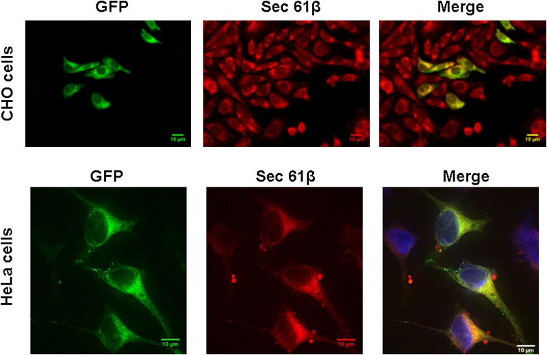
Fig. 2.
Localization of AGPAT11-GFP to ER in cultured cells. Upper panel: CHO cells overexpressing AGPAT11-GFP were fixed in methanol and incubated with antibody, specific for ER, sec61 and imaged for green and red fluorescence using fluorescence microscopy. Shown are representative images for sec61β (red fluorescence), AGPAT11 (green fluorescence), 4’-6-diamidino-2-phenylindole (blue fluorescence), and the merge. Lower panel: The HeLa cells were transiently transfected with AGPAT11-GFP construct. The cells were fixed as above after 48 h of transfection and processed as for CHO cells.

