Fig. 5.
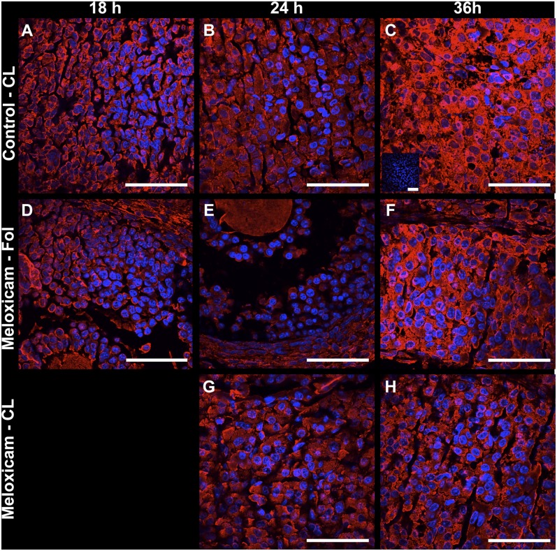
Protein expression of SCARB1 in representative corpora lutea (A–C, G, H) and in follicles (D–F) of immature mice stimulated with eCG and hCG and collected 18 h (A, D), 24 h(B, E, G) or 36 h after hCG (C, F, H). Mice received 0 (A–C) or 6 µg/g BW meloxicam, a prostaglandin synthase-2 activity inhibitor (D–H). Merged confocal microscopy images of fluorescent immunohistochemistry staining of SCARB1 and DAPI staining of nuclei. Panel C insert: An example of negative control (first antibody replaced by BSA). Bars: 50 µm. eCG, equine chorionic gonadotropin; hCG, human chorionic gonadotropin; SCARB1, scavenger receptor-B1.

