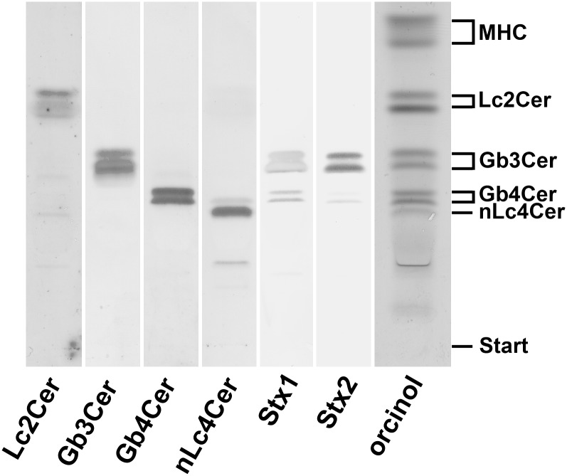
Fig. 1.
TLC immunodetection of individual GSLs in the neutral GSL preparation of human plasma. Amounts of GSLs applied for the orcinol stain correspond to 34.7 mg and those for the TLC overlay assays using anti-GSL antibodies as well as Stx1 and Stx2 are equivalent to 17.4 mg of human plasma proteins. Bound anti-GSL antibodies specific for Lc2Cer, Gb3Cer, Gb4Cer, and nLc4Cer as well as Stx1/anti-Stx1-antibody and Stx2/anti-Stx2-antibody were visualized with alkaline phosphatase conjugated secondary antibodies and BCIP as a substrate. The vertical white lines indicate areas of noncontiguous lanes assembled. The GSL structures and backgound information on the employed antibodies and Stxs are provided in Table 1. MHC, monohexosylceramides.

