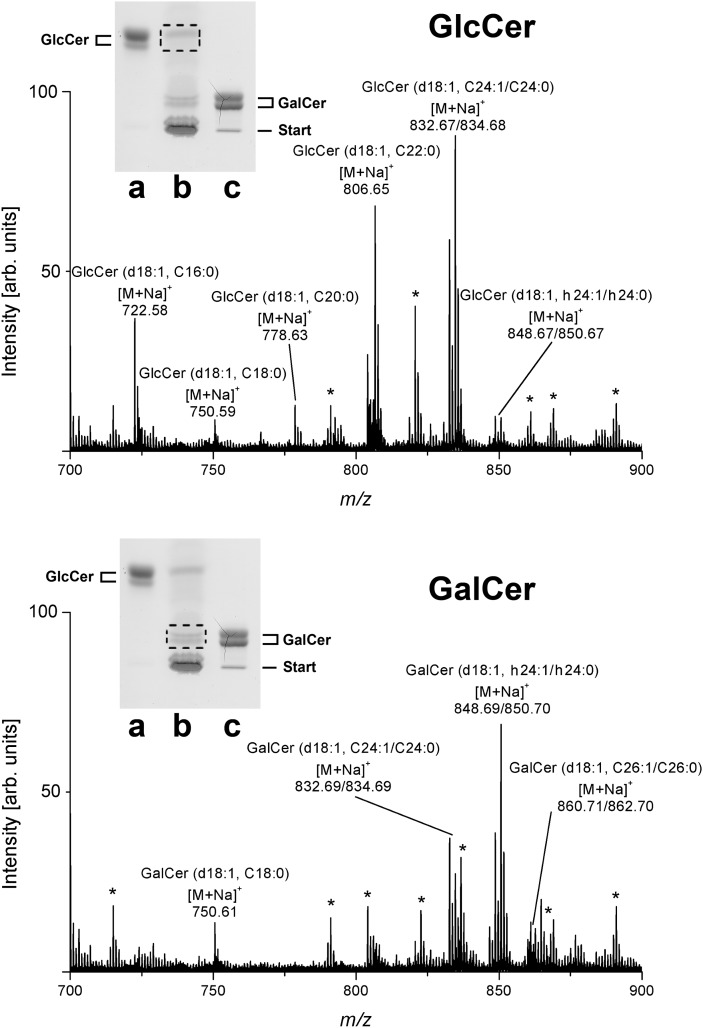
Fig. 2.
ESI Q-TOF-MS1 spectra of GlcCer and GalCer from human plasma. The spectra were prepared from silica gel extracts of primulin-stained monohexosylceramides after TLC separation as borate complexes in alkaline solvent. MS1 spectra of GlcCer (upper panel) and GalCer (lower panel) were recorded in the positive ion mode. Dotted frames in the insets, which show the orcinol stain of a TLC plate, indicate the silica gel positions from which primulin-detected GlcCer and GalCer were extracted, respectively. Lane a: Reference GlcCer, 7 µg; lane b: neutral GSL mixture corresponding to 34.7 mg of human plasma protein; lane c: reference GalCer, 10 µg. Unassigned ion species, which originate most likely from coextracted lipids are marked with asterisks and were not further analyzed. Major [M+Na]+ ions of monohexosylceramides and their proposed structures are listed in Table 2.

