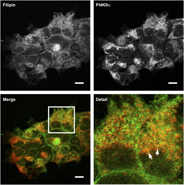
Fig. 3.
Distribution of sterol and endogenous PI4KIIα in A431 cells. Fixed cells were costained with filipin III (green channel) and an anti-PI4KIIα monoclonal antibody (red channel) and imaged by confocal microscopy. Arrows indicate membranes in the jutanuclear region with the greatest colocalization of filipin and PI4KIIα. Scale bars 10 μm. Images are representative of three independent experiments. PI4KII, type II phosphatidylinositol 4-kinase.

