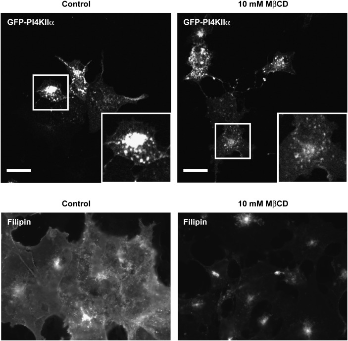
Fig. 4.
The effects of MβCD treatment on eGFP-PI4KIIα and sterol distribution. Cells expressing eGFP-PI4KIIα were treated with 10 mM MβCD for 20 min, fixed, and imaged by confocal microscopy. Boxed regions show detail of typical cells. Scale bar 20 μm. Cells treated with and without MβCD were also fixed and stained with filipin III and imaged by wide-field fluorescence microscopy to determine the extent of sterol depletion. Control images were collected to 12-bit peak, and identical gain settings were used in MβCD-treated cells. Images are representative of three independent experiments. eGFP, enhanced green fluorescent protein; MβCD, methyl-β-cyclodextrin; PI4KII, type II phosphatidylinositol 4-kinase.

