Fig. 5.
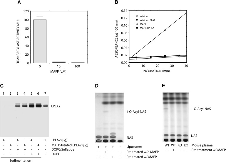
Effect of MAFP on LPLA2. A: Recombinant mouse LPLA2 was treated with 0, 10, or 100 µM MAFP for 1 h on ice. LPLA2 was incubated with DOPG/NAS (3:1, molar ratio) liposomes. The reaction mixture contained 130 µM phospholipid, 48 mM sodium citrate (pH 4.5), 10 µg/ml BSA, and 14.5 ng/ml of the enzyme in 500 µl of total volume and was incubated for 1.5–4.5 min at 37°C. The reaction products were extracted, separated, and quantified as described in Fig. 2. Error bars indicate SD (n = 3). B: Esterase activity of MAFP-treated LPLA2 was performed as described in Fig. 1. The reaction was initiated by adding vehicle or the enzyme and kept for 5, 15, 25, and 35 min at 37°C. C: Recombinant mouse LPLA2 (4 µg) pretreated with or without 100 µM MAFP as described in A was incubated with DOPC/ sulfatide-liposomes or DOPG-liposomes for 30 min on ice and centrifuged at 150,000 g for 1 h at 4°C. The resultant pellet was applied on SDS-PAGE as described in “Materials and Methods.” D and E: Human plasma (D) and mouse WT and LPLA2-knockout plasma (E) were pretreated with or without 100 µM MAFP for 1 h on ice. The treated plasma samples (2% for human and 4% for mice) were incubated with DOPG/NAS liposomes for 90 min at 37°C. The reaction products were analyzed by TLC as described in Fig. 1.

