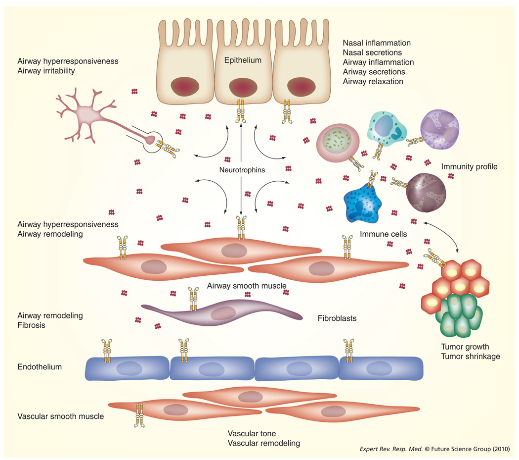
Figure 2. Neurotrophins in the lung.
As outlined in the main text, there is increasing evidence that NTs can be produced by, as well as targeted to, a number of lung components. Different NTs are expressed by nasal and airway epithelia, airway and vascular smooth muscle, sensory and other innervation of the lung, different immune cells, other structural cells and even tumors. As described in the article, NTs secreted by a cell type may have autocrine or paracrine effects on adjoining cell types. Thus NT signaling becomes central in the regulation of lung structure and function. In disease states (especially those involving inflammation), increased expression of NTs or their receptors can contribute to structural and functional changes such as enhanced inflammation, increased secretions, airway hyperresponsiveness (but impaired relaxation) and even altered tumor growth. See TABLE 1 for a summary of such effects involving specific NTs and their receptors.
NT: Neurotrophin.

