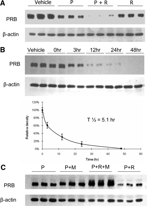
Figure 6.
Rosiglitazone treatment enhances ubiquitin-mediated PR degradation. A, Triplicate cultures of HEC-1A cells stably transfected with hPRB were treated with rosiglitazone, in the presence or absence of progesterone for 48 h. Cell lysates were probed with PR or β-actin antibodies by Western blot analysis. Note the profound reduction in PRB in presence of both agents. B, Duplicate cultures of HEC-1A cells stably transfected with hPRB were treated with progesterone and rosiglitazone for 48 h. Cell lysates were probed with PR and β-actin antibodies by Western blot analysis. The bottom graph shows the densitometric analysis of the ratio of PRB to β-actin at the indicated times. A half time for degradation was calculated to be 5.1 h. C, Triplicate cultures of HEC-1A cells stably transfected with hPRB were treated with rosiglitazone, progesterone, or MG 132 for 24 h as indicated. Cell lysates were probed with PR and β-actin antibodies by Western blot analysis. Note that inclusion of MG132 not only completely reverses the loss of PRB otherwise seen in the presence of rosiglitazone and progesterone but also results in accumulation of some larger PR forms presumably ubiquitinylated. Vehicle, 0.001% (vol/vol) ethanol + 0.1% (vol/vol) DMSO; P, 400 nm progesterone; R, 100 μm rosiglitazone; M, 100 μm MG132.

