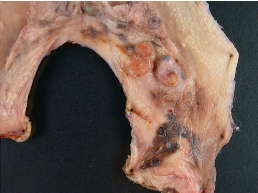
Figure 4.
This is a photograph of a primary vulvar melanoma. Grossly, the lesion shows variable pigmentation in an irregular distribution with focal polypoid tumor growth. Due to the irregular borders in this specimen it would be essential to diagram on a photograph or drawing the location from which sections are taken so that margins can be fully assessed and the exact location of any positive margins can be effectively communicated to the surgeon. Also important in this case is adequate sampling for measurement of maximal depth of invasion which will determine the pT for the melanoma.

