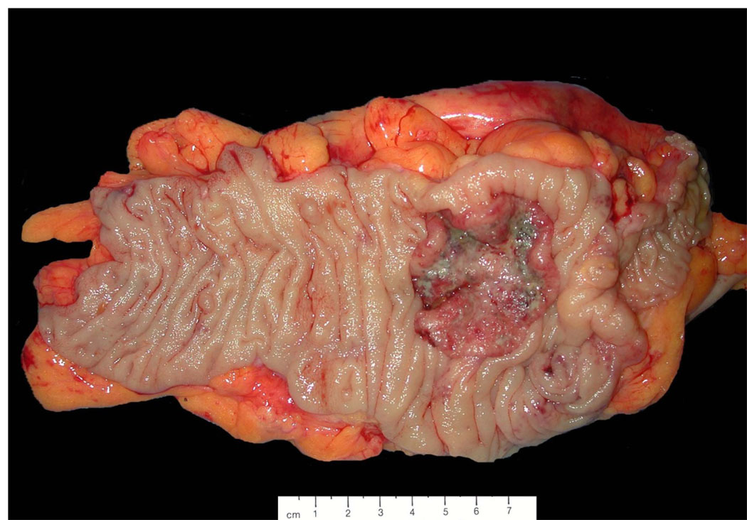
Figure 7.
Photograph of a primary cecal carcinoma. The specimen can be oriented based on gross identification of the terminal ilium (far right), ileocaecal valve, and appendix if present. Sections should be taken for histologic analysis to demonstrate the depth of invasion through the wall of the cecum (pT) and marginal status, and the associated adipose tissue should be carefully searched to find all lymph nodes for histologic examination (pN).

