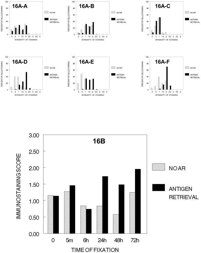
Figure 16.
DUI45 cells were either not fixed (0 min.) (16A-A) or fixed in 10% NBF at 4°C for 5 min. (16A–B), 6 hr. (16A–C), 24 hr. (16A–D), 48 hr. (16A–E) or 72 hr. (16A–F), washed, then all immersed in 70% ethanol for 1 hr., 80% ethanol for 1 hr., 90% ethanol for 1 hr., absolute ethanol for 3 hr., xylene for 2hr., followed by immersing in paraffin for 2 hr. The slides were then deparaffinized and rehydrated prior to antigen retrieval or no antigen retrieval and immunostaining to detect the PCNA antigen. Panel A demonstrates the percentage of cells stained at each intensity. Panel B shows the immunostaining score which was calculated as described.

