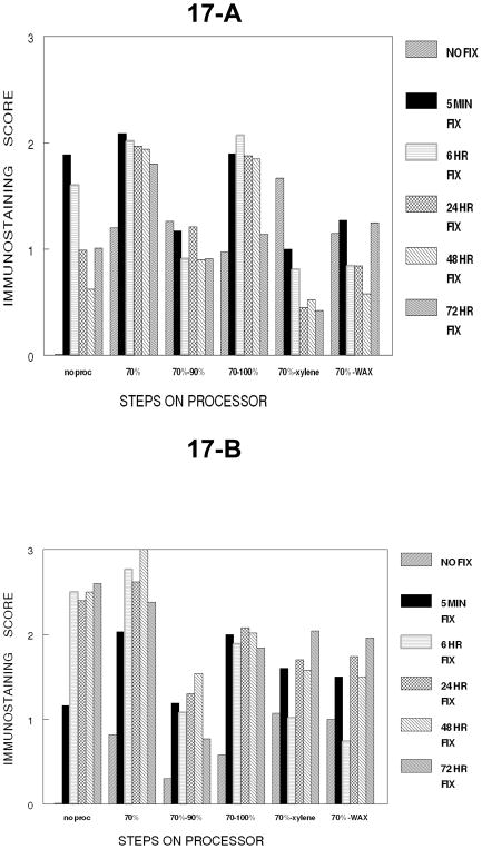
Figure 17.
Panel A of Figure 17 summarizes the immunostaining of PCNA in DU145 cells over all the cumulative steps of tissue processing. Note how fixation interacts with each cumulative step and that the transition to a hydrophobic environment has the greatest loss of immunorecognition for PCNA (p<0.001). Panel B shows the staining of PCNA following antigen retrieval for each of the cumulative steps of tissue processing at each time of fixation. Note that fixation for 24 hr. or greater gives the most consistent results for immunostaining for PCNA after antigen recovery.

