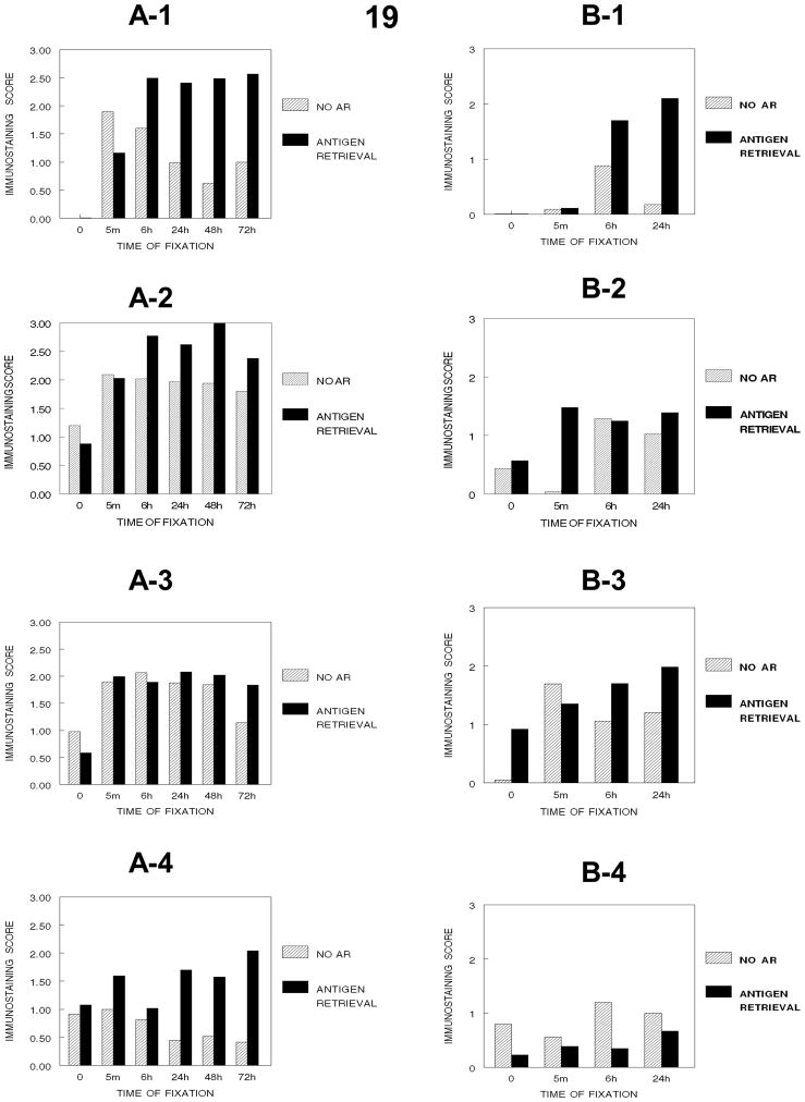
Figure 19.
Figure 19 demonstrates how immunorecognition of PCNA varies with the cumulative stages of tissue processing and the times of fixation in DU145 (A-1, no processing), (A-2, processing to 70% ethanol), (A-3, processing to absolute ethanol), (A-4, processing to xylene) and in SKOV-3 cells (B-1, no processing, B-2, processing to 70% ethanol, B-3, processing to absolute ethanol, B-4, processing to xylene). SKOV-3 cells were not evaluated beyond 24 hr. Note the major decrease in immunorecognition for PCNA occurs upon establishing a hydrophobic environment for DU145 cells (A-4) (p<0.001) and SKOV-3 cells (B-4) (p<0.001).

