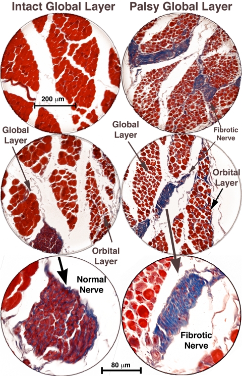
Figure 6.
Coronal histologic sections of the right and left orbits of monkey M1, 56 weeks after left ITN, showing marked atrophy of global layer fibers in the left SO muscle, with preservation of left orbital layer fibers. Fibrotic intramuscular nerves (shown at higher power in bottom panels) in the palsied left SO stain blue with Masson trichrome, whereas intact nerves in the right SO stain purple. Scale bars: (top two panels) 200 μm; (bottom panel) 80 μm.

