Figure 1.
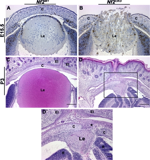
Nf2CKO lenses typically herniated through the area of lens-corneal fusion. (A) By E15.5, Nf2WT lenses show clear separation between the developing lens and the cornea. (B) Nf2CKO lenses usually developed lens herniation; fiber cells were expelled through the perforated cornea. (C) At P3, Nf2WT lenses were normal. (D, D′) Nf2CKO pups typically are born with abnormally small, disrupted lenses. Higher magnification shows the lens herniation phenotype. Expelled lens material is trapped between the cornea and the closed eyelids (asterisk). Sections in (A) and (B) were labeled with antibody to BrdU. Scale bars: 50 μm (A, B), 200 μm (C, D), 50 μm (D′, area highlighted by the black box in D). c, cornea; Le, lens; El, eyelid; R, retina.

