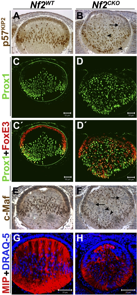
Figure 3.
Most Nf2CKO fiber cells express fiber-specific differentiation markers, but many coexpress epithelium-specific proteins. (A) At E13.5, high levels of p57KIP2 were present in nuclei at the lens equator and throughout the posterior fiber region of Nf2WT lenses. (B) In Nf2CKO lenses, many cells had weak expression or lacked detectable p57KIP2 (arrow and arrowheads). (C) In E12.5 Nf2WT lenses, antibodies to Prox1 labeled the nuclei of cells near the equator and throughout the posterior fiber region. (D) As in WT lenses, Prox1 staining was first detected in nuclei at the equator of Nf2CKO lenses. Careful examination revealed that all cells in the fiber region of the knockout lenses expressed Prox1, though levels were low in some cells. (C′) FoxE3 staining was present in the nuclei of all Nf2WT epithelial cells. Notably, several nuclei near the lens equator were stained with both antibodies. (D′) Antibodies to FoxE3 stained many cells in the posterior fiber compartment of these lenses. Therefore, all fiber cells in Nf2CKO lenses that expressed FoxE3 were also Prox1-positive. (E) In Nf2WT lenses, c-Maf is strongly expressed in the nuclei of all elongating fiber cells. (F) In Nf2CKO lenses, c-Maf is also localized to the nuclei of many fiber cells. However, there are many fiber cells that display either weak labeling or that lack positive labeling for c-Maf (arrowheads). This is especially evident in the clusters of cells near the equatorial region (arrow). Even when the images were contrast enhanced to give the impression of overstaining, the fiber cells in Nf2CKO lenses appeared unstained (arrow and arrowheads). (G, H) At E12.5, antibody to MIP stained fiber cells in Nf2WT and Nf2CKO lenses. However, not all Nf2CKO cells in the fiber compartment were stained. Cells in the epithelioid clusters near the equator had little or no detectable MIP staining. Scale bars: 25 μm (A, B, E, F), 50 μm (C, C′, D, D′), 20 μm (G, H).

