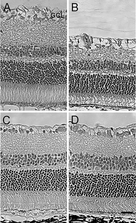
Figure 3.
Effect of TSA on retinal morphologic changes 7 days after unilateral ischemia. Animals were treated with vehicle or TSA (2.5 mg/kg) twice daily on days 0, 1, 2, and 3. (A, B) Photomicrographs of retinal cross-sections of contralateral and ischemic eyes from vehicle-treated animals. (C, D) Photomicrographs of retinal cross-sections of contralateral and ischemic eyes from TSA-treated animals. All micrographs were taken 2 to 3 disc diameters from the optic nerve. GCL, ganglion cell layer; INL, inner nuclear layer; ONL, outer nuclear layer. Scale bar, 20 mm.

