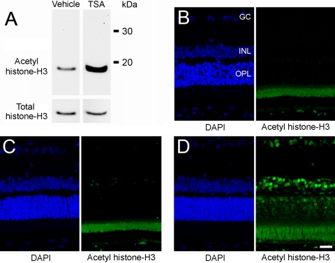
Figure 4.
Effect of TSA on retinal acetylation of histone-H3. Animals were treated with vehicle or TSA (2.5 mg/kg, administered intraperitoneally) 2 hours before tissue analysis. (A) Western blot analysis of retinal lysate for total and acetylated histone-H3. (B) DAPI staining of cell bodies and negative control (no primary antibody) for immunohistochemical staining of retinal acetylated histone-H3. Note the autofluorescence of the photoreceptor layer. (C) DAPI staining of cell bodies and immunohistochemical staining for retinal acetylated histone-H3 from vehicle-treated animals. (D) DAPI staining of cell bodies and immunohistochemical staining for retinal acetylated histone-H3 from TSA-treated animals. GC, ganglion cell layer; INL, inner nuclear layer; ONL, outer nuclear layer. Scale bar, 20 mm.

